


Gel Documentation System for DNA and RNA Analysis

Modern molecular biology depends on more than successful gel electrophoresis. Once DNA or RNA fragments have been separated, researchers need a reliable way to visualize, analyze, and preserve those results. This is where a gel documentation system for DNA and RNA analysis becomes an essential part of every laboratory workflow.
Whether you work in biotechnology, pharmaceutical research, academic laboratories, or quality control, accurate gel documentation supports confident decision-making and reproducible results. High-quality gel images help researchers compare samples, validate experiments, maintain laboratory records, and prepare publication-ready data.
In this guide, you'll learn how a gel documentation system works, why it matters for DNA and RNA analysis, and what features laboratories should consider when choosing the right solution.
What Is a Gel Documentation System?
A gel documentation system is a laboratory imaging instrument designed to capture high-resolution images of electrophoresis gels containing DNA, RNA, or proteins. After nucleic acid separation, the system uses controlled illumination, specialized filters, and a sensitive scientific camera to visualize stained bands with clarity.
Unlike conventional photography, a gel documentation system creates standardized images under consistent lighting conditions, making it easier to compare experiments and maintain reliable documentation.
The captured images can then be analyzed using dedicated software for band detection, image enhancement, and data storage, supporting research, diagnostics, and quality assurance.
Why Accurate DNA and RNA Documentation Matters
Every electrophoresis experiment generates valuable data. Proper documentation ensures those results remain useful long after the experiment is complete.
Accurate gel imaging helps laboratories:
- Improve reproducibility across repeated experiments.
- Maintain organized digital records for research and regulatory documentation
- Compare DNA and RNA band patterns with confidence.
- Prepare high-quality images for scientific publications and presentations.
- Support quality control in research and pharmaceutical laboratories.
Reliable imaging also minimizes interpretation errors by producing clear, consistent visualization of nucleic acid bands.
How a Gel Documentation System Works
The imaging process begins after electrophoresis, when stained DNA or RNA gels are placed inside the darkroom chamber of the imaging system.
A controlled illumination source, typically UV light or LED illumination, excites the fluorescent stain within the gel. A scientific CCD camera then captures detailed grayscale images through an optical filter that enhances band visibility while reducing unwanted background signals.
The accompanying analysis software allows users to acquire images, optimize brightness and contrast, archive results, and perform band analysis where required. This streamlined workflow enables laboratories to maintain consistency across experiments while reducing manual effort.
Features That Make a Difference in Gel Documentation
Not every gel imaging system offers the same level of performance. Laboratories often evaluate imaging quality, sensitivity, software capabilities, and ease of operation before making a purchase.
|
Feature |
Why It Matters |
|---|---|
|
Scientific CCD Camera |
Produces high-resolution gel images with excellent sensitivity. |
|
20 MP Resolution |
Captures fine DNA and RNA bands with greater clarity. |
|
16-bit Grayscale Imaging |
Provides detailed contrast for improved visualization. |
|
Motorized F1.2 Lens |
Enables precise focus and optimized image quality. |
|
UV Transilluminator (302 nm) |
Supports visualization of commonly used fluorescent stains. |
|
Optional White & Blue LED Illumination |
Expands compatibility with different staining methods. |
|
590 nm Filter |
Enhances gel documentation accuracy for supported applications. |
|
Integrated Analysis Software |
Simplifies image acquisition, storage, and analysis. |
|
Light-Tight Darkroom |
Ensures consistent imaging by eliminating external light interference. |
Applications of Gel Documentation Systems
A modern gel documentation system supports a wide range of molecular biology applications across research and industrial laboratories.
Its versatility makes it suitable for:
- DNA analysis for PCR products, restriction digestion, cloning, and genotyping.
- RNA analysis to evaluate extraction quality and integrity before downstream experiments.
- Protein gel documentation following electrophoresis using compatible staining methods.
- Research, biotechnology, pharmaceutical, and academic laboratories requiring reliable documentation and digital record keeping.
As laboratories continue to generate larger volumes of experimental data, dependable gel imaging has become an important part of efficient laboratory workflows.
Why Researchers Choose Althea Solutions Gel Documentation System
Selecting laboratory equipment is about more than specifications it is about reliability, consistency, and ease of use during everyday research.
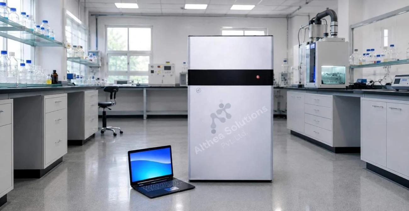
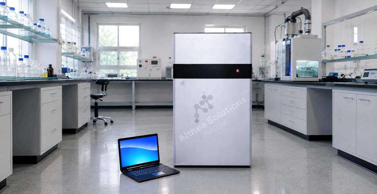
The Althea Solutions Gel Documentation System (ALH GDS-1520) is designed to support DNA, RNA, and protein gel imaging with a combination of sensitive optics, intelligent imaging, and practical laboratory features.
The system incorporates a 20 MP scientific CCD camera capable of producing detailed images with 16-bit grayscale output, helping laboratories visualize even low-intensity bands. Its F1.2 motorized lens with numeric focus and aperture adjustment enables consistent image acquisition, while the integrated autofocus function simplifies routine operation.
For nucleic acid imaging, the system includes a 302 nm UV transilluminator as standard and offers optional white LED and blue LED illumination for laboratories working with different staining chemistries. A 590 nm optical filter further enhances image quality for gel documentation applications.
The enclosed light-tight darkroom minimizes external light interference, while integrated gel documentation software supports image acquisition, storage, and analysis within a user-friendly workflow.
With compatibility for commonly used stains such as ethidium bromide, SYBR Gold, GelRed, GelStar, and SYPRO Orange, the system is suitable for diverse molecular biology applications across research laboratories, biotechnology organizations, universities, and pharmaceutical facilities.
Who Can Benefit from a Gel Documentation System?
The Althea Solutions Gel Documentation System is suitable for laboratories that require accurate and repeatable gel imaging as part of their daily workflow.
- Molecular biology laboratories
- Biotechnology companies
- Pharmaceutical quality control laboratories
- Universities and research institutions
- Clinical and diagnostic laboratories
- Contract research organizations (CROs)
- Academic teaching laboratories
Frequently Asked Questions
Q1. Is a gel documentation system only used for DNA analysis?
Answer: No. Modern systems support DNA, RNA, and protein gel imaging depending on the illumination source, filters, and staining methods used during electrophoresis.
Q2. Why is a scientific CCD camera important?
Answer: Scientific CCD cameras provide higher sensitivity, lower image noise, and improved grayscale detail compared to conventional cameras, allowing clearer visualization of nucleic acid bands.
Q3.Which stains are compatible with the Althea Solutions Gel Documentation System?
Answer: The system supports commonly used stains including Ethidium Bromide, SYBR Gold, GelRed, GelStar, and SYPRO Orange, depending on the selected illumination mode.
Q4. Can gel images be stored digitally?
Answer: Yes. Integrated gel documentation software enables laboratories to capture, organize, archive, and analyze digital gel images for future reference and reporting.
Q5. Which laboratories commonly use gel documentation systems?
Answer: They are widely used in molecular biology research, biotechnology, pharmaceutical development, diagnostic laboratories, academic institutions, and quality control laboratories.